Life Extension Magazine®
A serious and disabling disorder, osteoporosis is the most common metabolic bone disease in the Western world. Osteoporosis is characterized by low bone mass and the deterioration of bone structure leading to fragility and susceptibility to fracture. The most common fracture sites are the spine, hip, and forearm. Osteoporosis is more prevalent in women than in men, and its incidence increases with age. More than 10 million Americans have osteoporosis, and many more have a lesser form of weak bones called osteopenia.1 The health consequences of osteoporosis are far-reaching, as people who suffer a fracture due to osteoporosis have an increased risk of disability and mortality. The Dynamic State of BonesThroughout life, cells known as osteoblasts construct bone matrix and fill it with calcium. At the same time, cells called osteoclasts work just as busily to tear down and resorb the bone. This fine balance is regulated by many factors, including systemic hormones and cytokines. Bone mass reaches its peak by the middle of the third decade of life and plateaus for about 10 years, during which time bone turnover is constant, with bone formation approximately equaling bone resorption.2 As our bodies age, this fine balance is lost. As the relative hormone levels shift in midlife—more drastically in women than in men—the osteoclasts gain the upper hand and bone mass begins dwindling away. Some bone is already being lost by the time women reach menopause, but the rate of loss can increase as much as tenfold during the first six years after menopause. This is the essence of type I osteoporosis. From midlife onward, bone health is threatened by overactive osteoclasts. To add to the problem, the osteoblasts may become less active from age 60 onward, thus causing type II osteoporosis. Whereas trabecular (spongy-looking) bone in the vertebrae and elsewhere was formerly at risk from excess osteoclast activity, now the cortical (dense) bone of the hip, shin, pelvis, and other sites becomes more prone to fracturing because osteoblasts do not make enough of it. A fractured hip is an all-too-common prelude to death in elderly adults, and, at an average cost of $40,000, an economic burden on patients, families, and the health care system.3 Over 30% of all hip fractures occur in men.4 Who Is at Risk?Genetic factors help set the stage for osteoporosis.5 Americans of European or Asian ancestry are more susceptible than African-Americans. Our genetic “cards” are shuffled with each generation, so you may not have received all the same genes as your osteoporotic relative. Regardless of the genetic hand you were dealt, your chances of developing osteoporosis greatly depend on your environment and health habits. Individuals with a slight frame or a history of anorexia or bulimia are at greater risk for osteoporosis later in life. A history of amenorrhea, or the absence of menstrual periods, also increases risk. Smoking and heavy alcohol use promote weak, brittle bones. A high-phosphorus diet—epitomized by a fast-food hamburger and soft drink—causes a calcium-phosphorus imbalance that favors osteoporosis. Inactivity and lack of weight-bearing exercise, as in space travel or prolonged bed rest, also weaken the bones.
Age itself is a factor for bone health. Hormonal balance changes with age, as levels of bone-protecting sex hormones decline, more precipitously in women than in men. The hormonal form of vitamin D—1,25-dihydroxyvitamin D—declines, as do melatonin and dehydroepiandrosterone (DHEA). Cortisol and parathyroid hormone increase with age. Hormones such as testosterone, estrogen, and progesterone generally favor bone maintenance, as do vitamin D, DHEA, and melatonin.6 The thyroid, parathyroid, and glucocorticoid hormones (such as cortisol) favor bone destruction. Unfortunately, medications such as cortisone-type drugs cause some cases of osteoporosis. Antiepileptic drugs can also weaken the bones, apparently by interfering with vitamin D metabolism.7 Lithium, tamoxifen, and very high doses of thyroid hormone likewise may reduce bone mass.8 Reducing Risk with CalciumAdults can reduce their risk of osteoporosis by engaging in regular weight-bearing exercise, eating a variety of healthy foods, avoiding tobacco and alcohol, and minimizing their use of bone-weakening prescription drugs. Foods that promote bone health include calcium sources—such as dark green leafy vegetables, broccoli, legumes, canned salmon, and sardines—and dairy products like milk and cheese. Paradoxically, osteoporosis is quite common in Sweden and Norway, where milk consumption is high. Among the explanations advanced for this anomaly are excess vitamin A intake from fortified milk9 and widespread vitamin K deficiency.10 Daily calcium requirements vary by age. The National Academy of Sciences and National Osteoporosis Foundation recommend the following guidelines:11
While obtaining your daily calcium requirement from foods would be ideal, the fact is that many Americans’ diets often fall short in this regard. Hence, calcium supplements are good insurance against deficiency. Some doctors, no doubt attempting to be helpful, suggest chewable antacid tablets as a calcium supplement for their patients. That is not a good idea, however, because the type of calcium in those tablets—calcium carbonate—is poorly absorbed, especially in many elderly people who lack adequate stomach acid. Preventing deficiency should perhaps begin at an early age, when the bones are still growing. In one double-blind study, 94 teenage girls were given calcium citrate malate (a form of calcium preferred for its absorbability) or placebo for 18 months.12 The supplement increased the girls’ calcium intake from 80% of the recommended dietary allowance to 110%, with striking results: 24 grams more bone gained in the calcium group, amounting to an additional 1.3% of skeletal mass per year. This “head start” should protect these young women from the risk of osteoporotic bone fracture later in life.
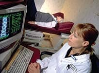
The same research team repeated their study of calcium citrate malate with a somewhat larger subject group for 24 months.13 The results and conclusions were similar. The calcium’s effectiveness depended on the subjects’ degree of physical maturity, a reflection of their sex hormone activity. Long after a person’s bone mass has begun to decline, calcium can still protect by slowing the rate of loss. In a study from the USDA’s Human Nutrition Research Center on Aging at Tufts University, men and women aged 65 or older were given 500 mg of calcium citrate malate and 700 IU of vitamin D daily for three years. The supplemented group had less than half as many non-vertebral fractures as the placebo group, and significantly less total-body bone loss.14 An earlier study from the same institution showed benefits from added calcium in postmenopausal women with inadequate calcium intakes; a supplement of calcium citrate malate performed better than calcium carbonate.15 Medical Tests and TreatmentsSeveral tests are available to measure bone mineral density, and all are considered good predictors of risk for future fracture. Dual-energy x-ray absorptiometry (DEXA) uses a small amount of radiation to measure bone density in the spine, hip, or wrist, the most common sites of osteoporotic fractures. Bone mineral density of the hip is considered the best predictor of hip fracture risk, while central DEXA of the hip and spine is the preferred measurement for definitive diagnosis. Peripheral DEXA or single-energy x-ray absorptiometry can be used to measure bone mineral density in the forearm, finger, and sometimes the heel. Quantitative computed tomography (QCT) measures trabecular and cortical bone density at several skeletal sites, but is most commonly used to measure trabecular bone density in the spine. Ultrasound densitometry assesses bone in the heel, tibia, patella, or other peripheral sites. While ultrasound measurements are generally not as precise as DEXA or QCT, they appear to be useful in predicting fracture risk.9 A diagnosis of osteoporosis is based on the measurement of bone mineral density at the spine, hip, or wrist. Normal bone density is indicated by a measurement that is within one standard deviation of a normal young adult, or a T-score between -1.0 and 1.0. The World Health Organization defines osteoporosis by a measurement of bone mineral density that is 2.5 standard deviations below that of a healthy young person, or a T-score at or below -2.5. T-scores between –1.0 and –2.5 signify osteopenia, a lesser degree of bone mineral loss.9
In addition, biochemical markers can be assessed to provide an indication of bone turnover. While these are useful for evaluating the success of therapy, they do not measure bone mass and so do not directly indicate the presence of osteoporosis. Biochemical markers include N-telopeptide, C-telopeptide, and deoxypyridinoline. Bone mineral density should be tested on all women aged 65 or older, in any postmenopausal woman with a fracture, and in postmenopausal women with one or more risk factors (such as smoking, low body weight, or use of certain prescription drugs). Some health providers recommend bone density screenings at an even younger age as part of a comprehensive approach to preventive health care. Medical treatments for osteoporosis include drugs, hormones, and nutrients.16 The most commonly used drugs are bisphosphonates and selective estrogen-response modifiers (SERMs). The bisphosphonates (such as Fosamax® and Actonel®) suppress osteoclast action, promote apoptosis (cell suicide) in osteoclasts, and prevent fractures. SERMs such as raloxifene (Evista®) provide the osteoclast-suppressing benefits of natural estrogen, without estrogen’s risk of uterine and breast cancer.
In place of SERMs, many integrative medical practitioners prefer soy isoflavones such as genistein and daidzein, which stimulate estrogen receptors in bone cells without undesirable activation of uterine and breast cells. In one recent double-blind, placebo-controlled trial in China, postmenopausal women received various amounts of soy extract along with conservative doses of calcium and vitamin D. The women who took the most soy (80 mg of isoflavones) lost the least bone from the hip area.17 The preponderance of osteoporosis in postmenopausal women, compared to men of similar age, was long believed to reflect estrogen deficiency. For many years, hormone replacement therapy was offered to women as an osteoporosis-prevention strategy, though most doctors prescribed horse-derived estrogens that are not identical to human estrogens. Because estrogen can promote breast and uterine cancers, and increase heart disease risk, its use is now controversial, with some clinicians recommending it18 and others warning against it.19 Male hormones such as testosterone build bone.20 In women, however, testosterone’s side effects include excess hairiness, or hirsutism, and deepening of the voice. The combination of testosterone and estrogen builds bone in osteoporotic women.21 The hormone calcitonin inhibits bone breakdown by osteoclasts. Calcitonin derived from salmon is available for administration by injection or as a nasal spray.2 Another hormone recently approved for medical use is teriparatide, which is a fragment of the human parathyroid hormone.22 The idea of using parathyroid hormone to treat osteoporosis might seem contraindicated, as sustained high levels of parathyroid hormone stimulate osteoclasts that resorb bone. However, when a patient is intermittently dosed with a synthesized fragment (the 1-34 part) of parathyroid hormone, the bone-building osteoblasts are stimulated.23 This therapy is an exciting new development because it can add bone mass.24,25 It works in conjunction with vitamin D,26 though combining it with a bisphosphonate seems to be unnecessary, or even detrimental.27 New therapies for the treatment of osteoporosis are under development. For example, Amgen is investigating the use of AMG 162, a genetically engineered antibody that mimics the action of the body’s natural osteoprotegerin protein for potential use in postmenopausal osteoporosis. Phase 3 studies on AMG 162 are currently under way. (More information is available at www.amgen.com.) | ||||||||||||||||||||
Nutritional TreatmentsOne treatment that can be considered both hormonal and nutritional is vitamin D. In its active, hormonal form, vitamin D enhances absorption of calcium from the small intestine. Its deficiency results in inadequate skeletal mineralization (rickets in children) or mineral depletion (osteomalacia in adults). When vitamin D is lacking, the parathyroid gland’s output increases to compensate for low calcium in the body, and the unfortunate result is more bone resorption—the hallmark of osteoporosis. Although 400-600 IU is the recommended daily dose of vitamin D for adults over the age of 50, the National Osteoporosis Found-ation recommends 800 IU for those at risk of deficiency. The safe upper dose is at least 2000 IU per day.28 Along with vitamin D, doctors routinely prescribe calcium for their osteoporotic patients and others at risk. One standard reference suggests a baseline preventive dietary intake of at least 1000 mg of calcium a day, unless osteoporosis risk factors or the disease itself is present, in which case the recommendation is 1000-1500 mg from calcium supplements.2 Because a daily dietary intake of 600 mg a day of calcium is about par for most Americans,3 calcium supplements clearly need to be used for preventing osteoporosis as well as treating it. Although everyone with osteoporosis can probably benefit from calcium, it is especially important for those on bisphosphonate drugs to receive adequate daily calcium and vitamin D. In fact, low blood calcium is a contraindication to the drugs.29 Many forms of supplemental calcium are available on the market. In one study, calcium in the form of its citrate and malate salts (calcium citrate malate) was absorbed better than milk calcium and calcium carbonate.30 In another study, calcium citrate malate was most absorbable, followed by milk calcium, calcium carbonate, and calcium phosphate.31 Calcium citrate decreases biochemical bone resorption markers unaffected by calcium carbonate.32 As for much-touted coral calcium, it outperformed only calcium carbonate in the sole comparison contained in the peer-reviewed literature.33 Calcium has many health benefits beyond maintaining bone strength. It helps regulate the heartbeat, transmits nerve impulses, assists blood clotting, activates enzymes, and stimulates hormone output. Calcium even protects against colon polyps, especially the kind most likely to turn cancerous.34
The Bone-Building TeamCalcium may excel at building bone, but it cannot work alone.35 Calcium’s bone-building team has many players. The most important are the mineral elements magnesium, manganese, boron, silicon, zinc, and copper, as well as vitamin D3, the natural form of vitamin D. Vitamins C and K also play key roles. The teamwork effect is nicely illustrated in a study by Strause and colleagues,36 who gave calcium citrate malate—either with or without the trace minerals zinc, manganese, and copper—to healthy postmenopausal women over a two-year period. The researchers evaluated spinal bone loss. The most loss occurred in women receiving placebo (with no calcium). At the end of the study, the group that had received both calcium citrate malate and the trace minerals was clearly better off than the placebo group, whereas other between-group comparisons were not significant. Magnesium is an important team player, with a role in regulating active calcium transport. Some studies have associated its dietary intake with increased bone mineral density. A two-year study of postmenopausal women supplemented with magnesium showed significant protection from osteoporosis and greater bone mineral density.37 Many doctors favor maintaining a magnesium intake that is approximately half your calcium intake as measured in milligrams.
Herbalists have long used a silicon-rich plant called horsetail to heal bones. In animals, silicon deficiency causes bone defects. Dietary silicon is absorbed as orthosilicic acid, which has recently been shown to stimulate the synthesis of collagen type 1 (the kind found in bone matrix) and to induce characteristic osteoblast enzymes in osteoblast-like cells.38 In a small retrospective study, women who received silica had significantly increased bone mineral density in the femur.39 Boron may mimic the action of estrogen, a hormone that protects bone health. In one report, postmenopausal women lost calcium and magnesium from their bodies when they were made boron deficient, and retained those minerals on a boron-supplemented diet; they also manufactured more estrogen and testosterone when on boron supplementation. In another study, vitamin D status improved in boron-deficient women after they received boron.40 Vitamin D acts as the “gatekeeper” of calcium by facilitating its absorption in the intestine. Natural vitamin D3 comes from sunshine and animal sources. A plant-derived form, vitamin D2 or ergocalciferol, is a common additive to dairy products. Vitamin D3, whether ingested or created naturally by the action of sunlight on the skin, is carried to internal organs that convert it into a potent, hormonal form. Only a few minutes of full sun exposure will create thousands of units of vitamin D3.41,42
If vitamin D is calcium’s gatekeeper in the body, vitamin K is its shepherd. Vitamin K ensures that calcium goes into the bones where it belongs, and stays out of parts of the body such as the arteries where it does not belong. It accomplishes this by helping an enzyme that activates special Gla-proteins that control calcium deposition.43 Using vitamin K does not require a physician’s supervision; however, because vitamin K is involved in blood clotting and may interact with certain medications, it should not be used by those taking anti-coagulant drugs like Coumadin®. Finally, vitamin C helps to create the collagen-rich bone matrix. One component of collagen (connective tissue) is hydroxyproline, an amino acid formed when proline becomes hydroxylated, a step that requires vitamin C. That is why bones break so easily and tissues are so weak in people with scurvy, which is the overt form of vitamin C deficiency. Although scurvy is rare, scientists like Linus Pauling and Irwin Stone long ago pointed out that most of us exist in a state of chronic subclinical scurvy due to near-universal insufficiency of vitamin C in our diet.44 Osteoporosis Prevention GuidelinesThe National Osteoporosis Foundation, which provides educational material to the public and health professionals, has posted on its website (www.nof.org) the following guidelines for preventing osteoporosis:
To these well-conceived rules, the Life Extension Foundation adds four more:
While a certain amount of bone loss may occur with aging, developing osteoporosis is certainly not inevitable. Regardless of genetic risks, most of us can maintain strong, fracture-resistant bones throughout our lifetimes by avoiding dietary and lifestyle habits that lead to bone loss, exercising regularly, receiving preventive health check-ups, and nourishing our bone-building cells with bioavailable calcium and other supportive nutrients. | ||||
| References | ||||
| 1. Available at: http://www.nof.org/advocacy/ prevalence. Accessed August 21, 2004. 2. Beers MH, Berkow R, eds. The Merck Manual, 17th ed. Whitehouse Station, NJ: Merck Research Laboratories; 1999:469-73. 3. Available at:.http://www.nof.org. Accessed August 21, 2004. 4. Seeman E. Osteoporosis in men. Baillieres Clin Rheumatol. 1997 Aug;11(3):613-29. 5. Baldock PA, Eisman JA. Genetic determinants of bone mass. Curr Opin Rheumatol. 2004 Jul;16(4):450-6. 6. Available at: http://www.gsdl.com. Accessed August 22, 2004. 7. Sandyk R, Anastasiadis PG, Anninos PA, Tsagas N. Is postmenopausal osteoporosis related to pineal gland functions? Int J Neurosci. 1992 Feb;62(3-4):215-25. 8. Elliott ME, Binkley N. Evaluation and mea- surement of bone mass. Epilepsy Behav. 2004 Feb;5 Suppl 2:S16-S23. 9. Available at: http://www.nof.org/physguide/diagnosis.htm. Accessed August 27, 2004. 10. Whiting SJ, Lemke B. Excess retinol intake may explain the high incidence of osteo- porosis in northern Europe. Nutr Rev. June 1999;57:192-5. 11. Drevon CA, Henriksen HB, Sanderud M, Gundersen TE, Blomhoff R. Biological effects of vitamin K and concentration of vitamin K in Norwegian food. Tidsskr Nor Laegeforen. 2004 Jun 17;124(12):1650-4. 12. Available at: http://www.nof.org/prevention/calcium.htm. Accessed August 26, 2004. 13. Lloyd T, Andon MB, Rollings N, et al. Calcium supplementation and bone mineral density in adolescent girls. JAMA. 1993 Aug 18;270(7):841-4. 14. Lloyd T, Martel JK, Rollings N, et al. The effect of calcium supplementation and Tanner stage on bone density, content and area in teenage women. Osteoporos Int. 1996;6(4):276-83. 15. Dawson-Hughes B, Dallal GE, Krall EA, Sadowski L, Sahyoun N, Tannenbaum S. A controlled trial of the effect of calcium sup- plementation on bone density in postmenopausal women. N Engl J Med. 1990 Sep 27;323(13):878-83. 16. Dawson-Hughes B, Harris SS, Krall EA, Dallal GE. Effect of calcium and vitamin D supplementation on bone density in men and women 65 years of age or older. N Engl J Med. 1997 Sep 4;337(10):670-6. 17. Available at: www.aeron.com. Accessed August 26, 2004. 18. Kirk D, Fish SA. Medical management of osteoporosis. Am J Manag Care. 2004 Jul;10(7 Pt 1):445-55. 19. Chen YM, Ho SC, Lam SS, Ho SS, Woo JL. Soy isoflavones have a favorable effect on bone loss in Chinese postmenopausal women with lower bone mass: a double-blind, randomized, controlled trial. J Clin Endocrinol Metab. 2003 Oct;88(10):4740-7. 20. Laszlo A. Postmenopausal osteoporosis. Orv Hetil. 2004 Jan;145(1):3-13. 21. Staren ED, Omer S. Hormone replacement therapy in postmenopausal women. Am J Surg. 2004 Aug;188(2):136-49. 22. Leifke E, Korner HC, Link TM, Berhre HM, Peters PE, Nieschlag E. Effects of testosterone replacement therapy on corti- cal and trabecular bone mineral density, vertebral body area, and paraspinal muscle area in hypogonadal men. Eur J Endocrinol. 1998 Jan;138(1):51-8. 23. Davis SR, McCloud P, Strauss BJ, Burger H. Testosterone enhances estradiol’s effects on postmenopausal bone density and sexu- ality. Maturitas. 1995 Apr;21(3):227-36. 24. Lee JR. Is natural progesterone the missing link in osteoporosis prevention and treatment? Med Hypotheses. 1991 Aug;35(4):316-8. 25. Whitfield JF, Morley P. Small bone-building fragments of parathyroid hormone: new therapeutic agents for osteoporosis. Trends Pharmacol Sci. 1995 Nov;16(11):382-6. 26. Frolik CA, Black EC, Cain RL, et al. Anabolic and catabolic bone effects of human parathyroid hormone (1-34) are predicted by duration of hormone exposure. Bone. 2003 Sep;33(3):372-9. 27. Quattrocchi E, Kourlas H. Teriparatide: a review. Clin Ther. 2004 Jun;26(6):841-54. 28. Lane NE. Parathyroid hormone: evolving therapeutic concepts. Curr Opin Rheumatol. 2004 Jul;16(4):457-63. 29. Slovik DM, Rosenthal DI, Doppelt SH, et al. Restoration of spinal bone in osteoporot- ic men by treatment with human parathyroid hormone (1-34) and 1,25-dihydroxyvita- min D. J Bone Miner Res. 1986 Aug;1(4):377-81. 30. Doggrell SA. Does the combination of alendronate and parathyroid hormone give a greater benefit than either agent alone in osteoporosis? Expert Opin Pharmacother. 2004 Apr;5(4):955-8. 31. Available at: http://www.nof.org/physguide/ univeral_recommendations.htm#adequate. Accessed August 27, 2004. 32. PDR, 58th ed. Montvale, NJ: Thomson PDR; 2004:1989, 2827-8. 33. Smith KT, Heaney RP, Flora L, Hinders SM. Calcium absorption from a new calcium delivery system (CCM). Calcif Tissue Int. 1987 Dec;41(6):351-2. 34. Cited in: PDR for Nutritional Supplements, 1st ed. Montvale, NJ: Medical Economics Co.; 2001:75. 35. Kenny AM, Prestwood KM, Biskup B, et al. Comparison of the effects of calcium loading with calcium citrate or calcium carbonate on bone turnover in postmenopausal women. Osteoporos Int. 2004 Apr;15(4):290- 4. 36. Ishitani K, Itakura E, Goto S, Esashi T. Calcium absorption from the ingestion of coral-derived calcium by humans. J Nutr Sci Vitaminol (Tokyo). 1999 Oct;45(5):509-17. 37. Wallace K, Baron JA, Cole BF, et al. Effect of calcium supplementation on the risk of large bowel polyps. J Natl Cancer Inst. 2004 Jun 16;96(12):921-5. 38. Tucker KL. Dietary intake and bone status with aging. Curr Pharm Des. 2003;9(32):2687-704. 39. Strause L, Saltman P, Smith KT, Bracker M, Andon MB. Spinal bone loss in post- menopausal women supplemented with calcium and trace minerals. J Nutr. 1994 Jul;124(7):1060-4. 40. PDR for Nutritional Supplements, 1st ed. Montvale, NJ: Medical Economics Co.; 2001:288-95. 41. Reffitt DM, Ogston N, Jugdaohsingh R, et al. Orthosilicic acid stimulates collagen type 1 synthesis and osteoblastic differentiation in human osteoblast-like cells in vitro. Bone. 2003 Feb;32(2):127-35. 42. Eisinger J, Clairet D. Effects of silicon, fluoride, etidronate and magnesium on bone mineral density: a retrospective study. Magnes Res. 1993 Sep;6(3):247-9. 43. PDR for Nutritional Supplements, 1st ed. Montvale, NJ: Medical Economics Co.; 2001:60-3. 44. Holick MF. Environmental factors that influence the cutaneous production of vitamin D. Am J Clin Nutr. 1995 Mar;61(3 Suppl):638S-45S. 45. Vieth R. Vitamin D supplementation, 25-hydroxyvitamin D concentrations, and safety. Am J Clin Nutr. 1999 May;69(5):842-56. 46. Shearer MJ. Role of vitamin K and Gla proteins in the pathophysiology of osteoporosis and vascular calcification. Curr Opin Clin Nutr Metab Care. 2000 Nov;3(6):433-8. 47. Stone I. Scurvy, the cosmic connection: an ancient supernova and the practice of medicine in the twentieth century. In: Huemer RP, ed. The Roots of Molecular Medicine: A Tribute to Linus Pauling. New York: W. H. Freeman; 1986:48-56. |

