Life Extension Magazine®

As we age, our soft tissues harden as a result of calcium infiltration. This calcification process is a major contributor to degenerative disease. Vitamin K functions to keep calcium out of soft tissues. New findings reveal that a vitamin K deficit creates more vascular calcification than initially thought.
A recent study showed when women were given a drug that blocks vitamin K, there was a 50% greater prevalence of arterial calcification compared to women not taking this drug (warfarin). This pathological effect occurred in as little as one month.1 This study showed that longer term use of warfarin was associated with even greater arterial calcification prevalence.
Kidney dialysis patients suffer severe arterial calcification with cardiovascular disease accounting for almost half of all their deaths.2 A clinical study examined vitamin K levels in dialysis patients. The results showed that 93% of the patients were at risk for arterial calcification. This risk was shown to be reduced with vitamin K2 supplementation.3
The quotation on the right side of this page comes from a report published by the American Heart Association.4 Despite this indisputable data, doctors today typically do nothing to protect their aging patients from the devastating impact of vascular calcification.
This article briefly describes how proper use of vitamin K can markedly protect our soft tissues against calcification.

“Most individuals aged over 60 years have progressively enlarging deposits of calcium mineral in their major arteries.5 This vascular calcification reduces aortic and arterial elastance, which impairs cardiovascular hemodynamics, resulting in substantial morbidity and mortality6-8 in the form of hypertension, aortic stenosis, cardiac hypertrophy, myocardial and lower-limb ischemia, congestive heart failure, and compromised structural integrity.9-11 The severity and extent of mineralization reflect atherosclerotic plaque burden12 and strongly and independently predict cardiovascular morbidity and mortality.”13
Source: American Heart Association
A search of the National Library of Medicine data base using the term “vascular calcification” at the time of this writing turns up the following numbers of published scientific articles:
Year New Articles
1982 16
1994 53
2004 214
2008 373
2014 700+
The total number of articles that discuss arterial calcification in the National Library of Medicine as of April, 2015, is over 6,400.
The surge from a mere 16 articles in 1982 to over 6,400 today is a reflection of the exponential increase in knowledge about this widespread pathological process.
The problem is that this lifesaving data is not being translated into clinical medical practice where it is urgently needed to protect aging humans against a host of cardio-vascular disorders.

Calcification And Hypertension
When arteries are soft and elastic, they readily expand and contract with each heartbeat. As arteries harden (calcify) and lose youthful elasticity, there is a progressive elevation in blood pressure.14
This happens because the heart is forced to beat stronger to force blood into the increasingly rigid arterial system. Calcification of the large artery exiting the heart (the aorta) helps explain why blood pressure elevates as people age.
A hallmark sign of long-term hypertension is enlargement of the heart’s left ventricle,15 which is the chamber of the heart that pushes blood into the aorta from where it is then distributed throughout the body.
The increase in cardiac workload caused by aortic rigidity (calcification) contributes to heart failure that afflicts over 5 million Americans.16,17
Aortic Valve Stenosis
A dilemma faced by elderly persons is progressive dysfunction of the valve between their heart and aorta that opens and closes with each heartbeat.
When the aortic valve fails to completely close, blood regurgitates back into the left ventricle of the heart.18,19 Without surgical replacement/repair of the aortic valve, death from congestive heart failure often occurs.20
Elderly persons are challenged to fully recover from aortic valve replacement, though newer intra-arterial techniques are becoming available whereby an artificial valve is threaded through the aorta and sewn into place.21 Those with successful mechanical valve replacements usually require anticoagulant drug therapy for life, which poses its own complicated set of side effects.22
It used to be thought that aortic stenosis was caused by a lifetime of “wear and tear.”23 It is now clear that calcification of the aortic valve leaflets is a cause of aortic valve failure, along with chronic inflammation, elevated glucose, high homocysteine, and low magnesium.24-31
Coronary Artery Calcification
Blockage of the coronary arteries that feed the heart muscle necessitates enormous amounts of hospital expenditures each year in the form of open heart coronary bypass surgeries and intra-arterial “stenting” procedures.
Interestingly, when open heart surgery is performed to replace a calcified aortic valve, the surgeon will often also bypass blocked coronary arteries in the same patient.32 This is not surprising since coronary atherosclerosis and aortic valve stenosis have similar underlying causes such as elevated homocysteine, chronic inflammation, and calcification.33-36
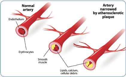
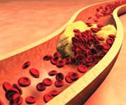
Calcification plays a significant role in accelerating the formation of the atherosclerotic plaque that narrows the coronary arteries of aging humans. In patients with coronary artery disease, calcification is present in 90% of cases.37,38
Clinically, vascular calcification is now accepted as a valuable predictor of coronary heart disease.39 Yet most cardiologists use it only as a diagnostic marker (the coronary calcium score test) as opposed to directly treating the underlying calcification pathology.40
What Causes Arteries To Calcify?

Many of the known risk factors that underlie atherosclerosis have been shown to promote arterial calcification. These include elevated LDL cholesterol, elevated homocysteine, diabetes, kidney failure, chronic inflammation, and oxidative stress.33,41-55
Additional calcification contributors include low magnesium (a natural calcium channel blocker), hormone imbalance, and excess blood calcium (caused by hyperparathyroidism).56-66
An underappreciated, major reason our vascular system turns to stone (calcifies) as we age, however, is inadequate intake of vitamin K.
As you’ll read next, a low blood level of vitamin K2 causes a protein in the vascular wall to bind calcium to arteries, heart valves, and other soft tissues.
A Calcium Inhibitor In Need Of Vitamin K
Matrix Gla-protein is a vitamin K-dependent protein, and it must be carboxylated to function properly. Poor vitamin K status leads to inactive uncarboxylated matrix Gla, which enables calcium to accumulate in soft tissues.67-69
Failure to optimally carboxylate matrix Gla-protein is a risk factor for atherosclerosis, coronary heart attack, and kidney disease.70-74 The title of a 2008 study that examined the impact of cardiovascular calcification is: “Matrix Gla-Protein: The Calcification Inhibitor In Need Of Vitamin K.”75,76
Matrix Gla-protein lines our vascular system and its function is governed by the quantity of vitamin K in our bloodstream.
When vitamin K levels are less than optimal, matrix Gla-protein allows calcium to infiltrate into our soft tissues similar to the way calcium absorbs into bone. When you hear the term “hardening of the arteries,” this can literally mean one’s previously flexible blood vessels are turning into rigid (calcified) bony structures.
With optimal levels of vitamin K, matrix Gla-protein becomes activated to shield calcium from entering arteries, heart valves, and other soft tissues.
Said differently, vitamin K functions as a control switch. When matrix Gla-protein is turned “on” by vitamin K, itblocks calcium from entering soft tissues. In the absence of adequate vitamin K, the matrix Gla-protein switch is turned “off” and calcium quickly infiltrates into soft tissues.
So the title of the 2008 study is quite revealing in that matrix Gla-protein is clearly a “ calcification inhibitor in need of vitamin K.”
What Is Matrix Gla-Protein?
 |
Early forms of life emanated from calcium-rich oceans, which required primitive organisms to develop mechanisms to prevent widespread calcium crystallization of living soft tissues.77
A prominent calcium-blocking mechanism is through activation of a protein called matrix gamma-carboxyglutamic acid, more commonly referred to as matrix Gla-protein or matrix Gla.78
The key to how matrix Gla-protein functions lies with its “carboxyl” group. Matrix Gla must be carboxylated to function properly.74
In the presence of vitamin K2, matrix Gla-protein becomes “carboxylated,” which means it’s being turned “ on” to repel calcium infiltration.74
Insufficient vitamin K2 results in matrix Gla being inadequately carboxylated or turned “off,” which means it’s unable to inhibit calcium infiltration into soft tissues.
To keep our natural calcium inhibitor matrix Gla continuously carboxylated, we need to provide it with a steady supply of vitamin K2. This is easy to do with one-per-day dosing of the proper forms of vitamin K.
What Happens When Vitamin K Is Acutely Withdrawn?
A recent study provided real-world evidence of what happens to aging humans who are deprived of vitamin K.
Warfarin is an anticoagulant drug that functions by antagonizing the effects of vitamin K in the body.79 This drug has been sold under the trade name Coumadin® for many decades.
Scientists were long ago aware that warfarin users suffered accelerated arterial calcification, but until recently, there was no alternative to protect high-risk patients from a thrombotic (arterial clotting) event, such as an ischemic stroke.
A study published in 2015 evaluated 451 women using mammograms to measure arterialcalcification. After just one month or more of warfarin drug therapy, the prevalence of arterial calcification increased by an astounding50% compared to that in untreated women. When these women were evaluated again after five years, the prevalence of arterial calcification increased almost 3-fold.1
This new human trial provides stark evidence of rapid calcification occurring in response to vitamin K withdrawal caused by warfarin (a vitamin K antagonist drug.)
We discuss the pros and cons of anticoagulant drugs that may be used in place of warfarin in this month’s issue of Life Extension® magazine.
What Happens When Vitamin K Is Introduced To Deficient Patients?
Kidney failure patients are kept alive by thrice weekly dialysis treatments. While the advent of hemodialysis has added countless human life years, it produces devastating side effects over the longer term. Over 50% of hemodialysis patients have vascular calcification, a major cause of cardiovascular disease. Cardiovascular disease accounts for about 50% of all deaths in these patients.80-82
A study was done to evaluate the effects of varying doses of the MK-7 form of vitamin K2 on markers of arterial calcification including carboxylation (activation) of matrix Gla-proteins.83
MK-7 (menaquinone-7) is a unique form of vitamin K2 because it remains active in the body for 24 hours and longer.84
At baseline, hemodialysis patients had a 4.5-fold higher level of uncarboxylated matrix Gla compared to controls.83 Daily doses of MK-7 of 45 mcg, 135 mcg, and 360 mcg were then administered over a six-week period.
Results were measured by the reduction of uncarboxylated matrix Gla and other measures of systemic calcification. Recall that when matrix Gla is under-carboxylated, it enables calcification of its surrounding tissue.
Supplementation with the MK-7 form of vitamin K2 reduced uncarboxylated matrix Gla by 36.7% in the 135 mcg dose group and 61.1% in the 360 mcg dose group. In the group given 360 mcg per day of MK-7, the favorable response rate was a remarkable 93%.83
When vitamin K2 supplementation was ceased in these dialysis patients, plasma levels of uncarboxylated matrix Gla-protein increased significantly, which indicated these high-risk individuals were once again vulnerable to severe vascular calcification.
Three Forms Of Vitamin K
 |
Based on the totality of evidence and low cost, it is prudent to take a supplement that contains multiple forms of vitamin K. The three forms of vitamin K most applicable to human health are:
- Vitamin K1 . Vitamin K1, also known as phylloquinone, is found in plants and some of it converts to vitamin K2 in the body.85 This form is considered the least effective because it depends on conversion into activated K2 to confer significant protection against calcification. There are nonetheless published studies showing disease risk reduction in response to ingestion of vitamin K1.86-90
- Vitamin K2 (MK-4) . MK-4 is found in meat, eggs, and dairy products.91 It is the most studied form of vitamin K to preserve bone health. It is rapidly absorbed and rapidly metabolized by the body.92-96
- Vitamin K2 (MK-7) . MK-7 is found in fermented soybeans and fermented cheeses.97,98 What makes this form so special is that it remains active in the body for more than 24 hours.84 This is critical when protecting against calcification since matrix Gla-proteins quickly inactivate in the absence of vitamin K2.99
The federal government says that adults only need 90 to 120 mcg per day of vitamin K.100-102 While this is enough to enable proper blood clotting, current research suggests it falls below levels needed to protect against vascular calcification.99,103-106
Importance Of Adequate Calcium Intake
Calcium serves numerous life-sustaining processes, the most important of which is to maintain the electrolyte balance needed for proper rhythmic heart beats.107 If one were to deplete their bloodstream of calcium, they could die from a heart attack caused by an acute arrhythmic disorder.
In a healthy body, 99% of all calcium is stored in bone where it provides structural support.108 The amount of calcium that is allowed in the bloodstream is tightly controlled by the parathyroid glands.109
In bone, vitamin K2 activates proteins that bind calcium.110 Populations with high dietary intake of vitamin K2 have lower rates of osteoporosis.111-114
People need around 1,200 mg per day of calcium from diet and supplements to maintain bone density. We suggest around 700 mg per day of supplemental calcium for women and around 500 mg per day for men. Most people can rely on their diet for the balance of their calcium needs.115
Supplementing with moderate daily doses of calcium will not accelerate arterial calcification.116,117 One reason is that calcium blood levels are tightly regulated in the body and most ingested calcium will be stored in one’s bones. Your bloodstream has top priority when it comes to getting the calcium it needs, which means that if you intentionally deprive yourself of calcium from food and supplements, your parathyroid glands will rob calcium from your bones to maintain a constant calcium blood level to ensure electrolyte balance.118
Vitamin K is a calcium-regulating nutrient. When properly supplemented, vitamin K2 activates matrix Gla-proteins in soft tissues to keep calcium out. On the flip side, vitamin K2 activates calcium binding proteins in bone to maintain skeletal density.
In the absence of vitamin K, bony structures form in soft tissues. Early pathologists were perplexed to find arteries that were supposed to be soft and pliable instead had literally turned to stone. In 1863, Rudolf Virchow, known as the “father of pathology,” described vascular changes he observed as “ossification, not mere calcification, occurring by the same mechanism by which an osteophyte forms on the surface of bone.” 119
These observations confirmed by modern findings clearly demonstrate the power of vitamin K, or lack thereof, to control whether we maintain strong bone density and soft pliable tissues, or develop osteoporosis together with vascular calcification.
Can Calcification Be Reversed?
Most adults probably suffer some degree of calcification, as intake of vitamin K in Western societies remains at epidemic low levels.
Some of us are severely calcified because of medical disorders requiring dialysis or the drug warfarin, or we allowed blood levels of homocysteine, LDL, or glucose to remain too high for too long.
So the question begs is there anything we can do now to reverse the accumulation of calcium in our arteries, heart valves, glands, and other soft tissues? We found one animal study published in 2007 suggesting that high-dose vitamin K might work. The authors of the study wrote: 99
“Given that arterial calcifications are predictive of cardiovascular events, regression of arterial calcification may help to reduce the risk of death in people with chronic kidney disease and coronary artery disease.”
The study involved four groups of rats who were all initially fed a six-week diet that contained warfarin to induce calcium buildup in the blood vessels. This diet also included a low-dose (normal) vitamin K1 to ensure the animals were not vitamin K deficient. The rats were divided into several groups, of which the following four groups comprised the main part of the experiment:

Group 1: Continue thewarfarin plus normal K1 diet;
Group 2: Stop warfarin, but continue with normal dose of vitamin K1;
Group 3: Stop warfarin, but use a high-dose of vitamin K1;
Group 4: Stop warfarin, but add high-dose of MK-4 formof vitamin K2.
During the initial six weeks of warfarin plus normal K1, all animals showed a significant increase in arterial calcification.
In the groups receiving high-dose vitamin K1 or K2 (MK-4), not only was there no further arterial calcium accumulation, but there was a greater than 37% reduction of previously accumulated arterial calcification after six weeks. After 12 weeks, there was a 53% reduction in accumulated arterial calcium deposits.
The groups receiving the high-dose vitamin K1 and K2 also showed a reversal in carotid artery stiffness.99
This study provides intriguing evidence that warfarin-induced calcification may be reversible by high vitamin K intake.
An estimate of the human equivalent dose given to the rats whose arterial calcification was reversed is difficult to precisely calculate because of many variables involved. Our calculations based on estimates of food consumption and animal body weight suggest that the human equivalent dose of the vitamin K2 (MK-4) used in this study is in the range of approximately 52,000 mcg to 97,000 mcg per day (i.e. 52 mg to 97 mg per day).
Since the RDA for vitamin K is only 90 to 120 mcg, the dose of vitamin K used in this rat study may seem extremely high. Yet in Japan, the MK-4 form of vitamin K2 is approved as a drug to treat osteoporosis in humans, and the daily dose is 45,000 mcg (45 mg), which has not been reported to have any toxic effects.120-123
Most Life Extension® members take a combination supplement that provides 1,000 mcg of vitamin K1, 1,000 mcg of vitamin K2 (MK-4), and 200 mcg of vitamin K2 (MK-7).
These are high doses compared to dietary supplement industry standards, and one could postulate that taking these daily doses over an extended time period might induce a regression of arterial calcification, but more human research is needed to establish this. Some members are taking higher doses of vitamin K now with the objective of reversing accumulated soft tissue calcification.
 |
Systemic Calcification
Cardiovascular tissues are particularly prone to calcium infiltration.
The calcification process, however, is also commonly observed in the skin, kidney, tendons, glands, and other soft tissues as a result of disease, and/or aging.124-132
How To Properly Supplement With Vitamin K
A review of the published scientific literature provides a rationale for aging people to supplement with all three vitamin K forms, i.e. vitamin K1, vitamin K2 (MK-4), and vitamin K2 (MK-7).
Since vitamin K is fat-soluble, taking it with the fattiest meal of the day will greatly augment absorption into one’s bloodstream.
A lot of members ask why they cannot take just the MK-7 form of vitamin K2 since this has long-acting effects in the body and has demonstrated powerful calcium blocking properties. Our response is that vitamin K1 and MK-4 have demonstrated impressive results in other studies, so it is best to take a formula that contains all three forms of vitamin K. As mentioned already, the MK-4 form of vitamin K2 has been used in high doses as a prescription drug in Japan to treat osteoporosis.
Since vitamin K1 and MK-4 are inexpensive, it makes sense to include them with the long-acting MK-7 form of vitamin K2 to inhibit and possibly reverse as much vascular calcification as possible, while providing support for strong bones.
Why Doctors Are Apprehensive About Vitamin K
In 1999, one of our scientific advisors recommended to me that aging people supplement with vitamin K.
Vitamin K is known as the “coagulation vitamin” because of the critical role in plays in essential blood clotting.
I was initially apprehensive because abnormal clotting inside blood vessels (thrombosis) is a leading cause of death in the elderly. 133,134 Thrombosis is a frequent underlying cause of coronary occlusion heart attacks and ischemic strokes.135,136
One might think that taking higher amounts of vitamin K would increase thrombotic risk. This concern, however, has no basis in reality. The reason
is that only small amounts of vitamin K are required to fully saturate
coagulation
proteins
.137
Once coagulation proteins are fully saturated by vitamin K, then there is no increased thrombotic risk in response to additional vitamin K intake.
The misconception about the role vitamin K plays in coagulation is contributing to the epidemic of diseases caused by vascular calcification. A plethora of published studies indicate that the most common degenerative diseases afflicting aging humans could be prevented by taking the proper doses of vitamin K.
What mainstream doctors still don’t understand is vitamin K’s critical role of blocking the calcification of heart valves, arterial linings, and other soft tissues, while helping to keep calcium in bone where it is needed.
Eye-opening recent studies shed light on the importance for people to optimize their intake of vitamin K to protect against soft tissue calcification.
An urgent need exists to convey this information to the medical community. This may not happen any time soon because vitamin K is sold as a low-cost dietary supplement and not an expensive prescription drug.
For longer life,
William Faloon
References
- Tantisattamo E, Han KH, O’Neill WC. Increased vascular calcification in patients receiving warfarin. Arterioscler Thromb Vasc Biol. 2015 Jan;35(1):237-42.
- Salusky IB, Goodman WG. Cardiovascular calcification in end-stage renal disease. Nephrol Dial Transplant. 2002 Feb;17(2):336-9.
- Pilkey RM, Morton AR, Boffa MB, et al. Subclinical vitamin K deficiency in hemodialysis patients. Am J Kidney Dis. 2007 Mar;49(3):432-9.
- Allison MA, Criqui MH, Wright CM. Patterns and risk factors for systemic calcified atherosclerosis. Arterioscler Thromb Vasc Biol. 2004 Feb;24(2):331-6.
- Demer LL. Cholesterol in vascular and valvular calcification. Circulation. 2001 Oct 16;104(16):1881-3.
- Wayhs R, Zelinger A, Raggi P. High coronary artery calcium scores pose an extremely elevated risk for hard events. J Am Coll Cardiol. 2002 Jan 16;39(2):225-30.
- Arad Y, Spadaro LA, Goodman K, Newstein D, Guerci AD. Prediction of coronary events with electron beam computed tomography. J Am Coll Cardiol. 2000 Oct;36(4):1253-60.
- Keelan PC, Bielak LF, Ashai K, et al. Long-term prognostic value of coronary calcification detected by electron-beam computed tomography in patients undergoing coronary angiography. Circulation. 2001 Jul 24;104(4):412-7.
- Kelly RP, Tunin R, Kass DA. Effect of reduced aortic compliance on cardiac efficiency and contractile function of in situ canine left ventricle. Circ Res.1992 Sep;71(3):490-502.
- Ohtsuka S, Kakihana M, Watanabe H, Sugishita Y. Chronically decreased aortic distensibility causes deterioration of coronary perfusion during increased left ventricular contraction. J Am Coll Cardiol. 1994 Nov 1;24(5):1406-14.
- Watanabe H, Ohtsuka S, Kakihana M, Sugishita Y. Decreased aortic compliance aggravates subendocardial ischaemia in dogs with stenosed coronary artery. Cardiovasc Res. 1992 Dec;26(12):1212-8.
- Sangiorgi G, Rumberger JA, Severson A, et al. Arterial calcification and not lumen stenosis is highly correlated with atherosclerotic plaque burden in humans: a histologic study of 723 coronary artery segments using nondecalcifying methodology. J Am Coll Cardiol. 1998 Jan;31(1):126-33.
- Vliegenthart R, Oudkerk M, Hofman A, Oei HH, van Dijck W, van Rooij FJ, Witteman JC. Coronary calcification improves cardiovascular risk prediction in the elderly. Circulation. 2005 Jul 26;112(4):572-7.
- Kalra SS, Shanahan CM. Vascular calcification and hypertension: cause and effect. Ann Med. 2012 Jun;44 Suppl 1:S85-92.
- Messerli FH, Aepfelbacher FC. Hypertension and left-ventricular hypertrophy. Cardiol Clin. 1995 Nov;13(4):549-57.
- Leening MJ, Elias-Smale SE, Kavousi M, et al. Coronary calcification and the risk of heart failure in the elderly: the Rotterdam Study. JACC Cardiovasc Imaging. 2012 Sep;5(9):874-80.
- Available at: http://www.cdc.gov/dhdsp/data_statistics/fact_sheets/fs_heart_failure.htm. Accessed February 12, 2015.
- Available at: http://www.ncbi.nlm.nih.gov/pubmedhealth/pmh0062945. Accessed April 17, 2015.
- Bekeredjian R, Grayburn PA. Valvular heart disease: aortic regurgitation. Circulation. 2005 Jul 5;112(1):125-34. Review. Erratum in: Circulation. 2005 Aug 30;112(9):e124.
- Dujardin KS, Enriquez-Sarano M, Schaff HV, Bailey KR, Seward JB, Tajik AJ. Mortality and morbidity of aortic regurgitation in clinical practice. A long-term follow-up study. Circulation. 1999 Apr 13;99(14):1851-7.
- Webb JG, Pasupati S, Humphries K, et al. Percutaneous transarterial aortic valve replacement in selected high-risk patients with aortic stenosis. Circulation. 2007 Aug 14;116(7):755-63.
- Williams JW Jr, Coeytaux R, Wang A, et al. Percutaneous heart valve replacement. Comparative Effectiveness Technical Briefs, No. 2. Rockville (MD): Agency for Healthcare Research and Quality (US); 2010 Aug. Introduction.
- Novaro GM, Griffin BP. Calcific aortic stenosis: another face of atherosclerosis? Cleve Clin J Med. 2003 May;70(5):471-7.
- Dweck MR, Jones C, Joshi NV, et al. Assessment of valvular calcification and inflammation by positron emission tomography in patients with aortic stenosis. Circulation. 2012 Jan 3;125(1):76-86.
- Mohty D, Pibarot P, Després JP, et al. Association between plasma LDL particle size, valvular accumulation of oxidized LDL, and inflammation in patients with aortic stenosis. Arterioscler Thromb Vasc Biol. 2008 Jan;28(1):187-93.
- Latsios G, Tousoulis D, Androulakis E, et al. Monitoring calcific aortic valve disease: the role of biomarkers. Curr Med Chem. 2012;19(16):2548-54.
- Paolisso G, Tagliamonte MR, Rizzo MR, et al. Prognostic importance of insulin-mediated glucose uptake in aged patients with congestive heart failure secondary to mitral and/or aortic valve disease. Am J Cardiol. 1999 May 1;83(9):1338-44.
- Katz R, Wong ND, Kronmal R, et al. Features of the metabolic syndrome and diabetes mellitus as predictors of aortic valve calcification in the Multi-Ethnic Study of Atherosclerosis. Circulation. 2006 May 2;113(17):2113-9.
- Novaro GM, Aronow HD, Mayer-Sabik E, Griffin BP. Plasma homocysteine and calcific aortic valve disease. Heart. 2004 Jul;90(7):802-3.
- Gunduz H, Arinc H, Tamer A, et al. The relation between homocysteine and calcific aortic valve stenosis. Cardiology. 2005 103(4):207-11.
- Hruby A, O’Donnell CJ, Jacques PF, Meigs JB, Hoffmann U, McKeown NM. Magnesium intake is inversely associated with coronary artery calcification: the Framingham Heart Study. JACC Cardiovasc Imaging. 2014 Jan;7(1):59-69.
- Kobayashi KJ, Williams JA, Nwakanma L, Gott VL, Baumgartner WA, Conte JV. Aortic valve replacement and concomitant coronary artery bypass: assessing the impact of multiple grafts. Ann Thorac Surg. 2007 Mar;83(3):969-78.
- Sainani GS, Sainani R. Homocysteine and its role in the pathogenesis of atherosclerotic vascular disease. J Assoc Physicians India. 2002 May;50 Suppl:16-23.
- Wilson PW. Evidence of systemic inflammation and estimation of coronary artery disease risk: a population perspective. Am J Med. 2008 Oct;121(10 Suppl 1):S15-20.
- Budoff MJ. Atherosclerosis imaging and calcified plaque: coronary artery disease risk assessment. Prog Cardiovasc Dis. 2003 Sep-Oct;46(2):135-48.
- Available at: http://www.nhlbi.nih.gov/health/health-topics/topics/hs/types. Accessed April 18, 2015.
- Liu YC, Sun Z, Tsay PK, Chan T, Hsieh IC, Chen CC, Wen MS, Wan YL. Significance of coronary calcification for prediction of coronary artery disease and cardiac events based on 64-slice coronary computed tomography angiography. Biomed Res Int. 2013;2013:472347.
- Eisen A, Tenenbaum A, Koren-Morag N, et al. Coronary and aortic calcifications inter-relationship in stable angina pectoris: A Coronary Disease Trial Investigating Outcome with Nifedipine GITS (ACTION)--Israeli spiral computed tomography substudy. Isr Med Assoc J. 2007 Apr;9(4):277-80.
- Greenland P, Bonow RO, Brundage BH, et al. ACCF/AHA 2007 clinical expert consensus document on coronary artery calcium scoring by computed tomography in global cardiovascular risk assessment and in evaluation of patients with chest pain: a report of the American College of Cardiology Foundation Clinical Expert Consensus Task Force. Circulation. 2007 115:402-26.
- Fernandez-Friera L, Garcia-Alvarez A, Bagheriannejad-Esfahani F, et al. Diagnostic value of coronary artery calcium scoring in low-intermediate risk patients evaluated in the emergency department for acute coronary syndrome. Am J Cardiol. 2011 Jan;107(1):17-23.
- Van Campenhout A, Moran CS, Parr A, et al. Role of homocysteine in aortic calcification and osteogenic cell differentiation. Atherosclerosis. 2009 Feb;202(2):557-66.
- Allison MA, Wright M, Tiefenbrun J. The predictive power of low-density lipoprotein cholesterol for coronary calcification. Int J Cardiol. 2003 Aug;90(2-3):281-9.
- Kroon PA. Cholesterol and atherosclerosis. Aust N Z J Med. 1997. Aug;27(4):492-6.
- Jialal I, Devaraj S. Low-density lipoprotein oxidation, antioxidants, and atherosclerosis: a clinical biochemistry perspective. Clin Chem. 1996 Apr;42(4):498-506.
- Wilson PW, D’Agostino RB, Levy D, Belanger AM, Silbershatz H, Kannel WB. Prediction of coronary heart disease using risk factor categories. Circulation. 1998 May 12;97(18):1837-47.
- Liabeuf S, Olivier B, Vemeer C, et al. Vascular calcification in patients with type 2 diabetes: the involvement of matrix Gla-protein. Cardiovasc Diabetol. 2014 Apr 24;13:85.
- Chen NX, Moe SM. Arterial calcification in diabetes. Curr Diab Rep. 2003 Feb;3(1):28-32.
- Reaven PD, Sacks J. Coronary artery and abdominal aortic calcification are associated with cardiovascular disease in type 2 diabetes. Diabetologia . 2005 Feb;48(2):379-85.
- Niskanen LK, Suhonen M, Siitonen O, Lehtinen JM, Uusitupa MI. Aortic and lower limb artery calcification in type 2 (non-insulin-dependent) diabetic patients and non-diabetic control subjects. A five year follow-up study. Atherosclerosis. 1990 Sep;84(1):61-71.
- Goodman WG, London G, Amann K, et al. Vascular calcification in chronic kidney disease. Am J Kidney Dis. 2004 Mar;43(3):572-9. Review.
- Sharon M. Moe, Neal X. Chen. Mechanisms of vascular calcification in chronic kidney disease. JASN. 2008 Feb;(19)2:213-6.
- Qunibi WY. Reducing the burden of cardiovascular calcification in patients with chronic kidney disease. J Am Soc Nephrol. 2005 16(suppl 2):S95-S102.
- Doherty TM, Asotra K, Fitzpatrick LA, et al. Calcification in atherosclerosis: bone biology and chronic inflammation at the arterial crossroads. Proc Natl Acad Sci U S A. 2003 Sep 30;100(20):11201-6
- Nadra I, Mason JC, Philippidis P, et al. Proinflammatory activation of macrophages by basic calcium phosphate crystals via protein kinase C and MAP kinase pathways: a vicious cycle of inflammation and arterial calcification? Circ Res. 2005 Jun 24;96(12):1248-56.
- Byon CH, Javed A, Dai Q, et al. Oxidative stress induces vascular calcification through modulation of the osteogenic transcription factor Runx2 by AKT signaling. J Biol Chem. 2008 May 30;283(22):15319-27.
- Del Gobbo LC, Imamura F, Wu JH, de Oliveira Otto MC, Chiuve SE, Mozaffarian D. Circulating and dietary magnesium and risk of cardiovascular disease: a systematic review and meta-analysis of prospective studies. Am J Clin Nutr. 2013 Jul;98(1):160-73.
- Gorgels TG, Waarsing JH, de Wolf A, ten Brink JB, Loves WJ, Bergen AA. Dietary magnesium, not calcium, prevents vascular calcification in a mouse model for pseudoxanthoma elasticum. J Mol Med (Berl). 2010 May;88(5):467-75.
- van den Broek FA, Beynen AC. The influence of dietary phosphorus and magnesium concentrations on the calcium content of heart and kidneys of DBA/2 and NMRI mice. Lab Anim. 1998 Oct;32(4):483-91.
- Michos ED, Vaidya D, Gapstur SM, et al. Sex hormones, sex hormone binding globulin, and abdominal aortic calcification in women and men in the multi-ethnic study of atherosclerosis (MESA). Atherosclerosis. 2008 Oct;200(2):432-8.
- Joffe HV, Ridker PM, Manson JE, Cook NR, Buring JE, Rexrode KM. Sex hormone-binding globulin and serum testosterone are inversely associated with C-reactive protein levels in postmenopausal women at high risk for cardiovascular disease. Ann Epidemiol. 2006 Feb;16(2):105-12.
- Sarkar NN. Hormonal profiles behind the heart of a man. Cardiol J. 2009 16(4):300-6.
- Calderon-Margalit R, Schwartz SM, Wellons MF, et al. Prospective association of serum androgens and sex hormone-binding globulin with subclinical cardiovascular disease in young adult women: the Coronary Artery Risk Development in Young Adults women’s study. J Clin Endocrinol Metab. 2010 Sep;95(9):4424-31.
- Tordjman KM, Yaron M, Izkhakov E, et al. Cardiovascular risk factors and arterial rigidity are similar in asymptomatic normocalcemic and hypercalcemic primary hyperparathyroidism. Eur J Endocrinol. 2010 May;162(5):925-33.
- Han D, Trooskin S, Wang X. Prevalence of cardiovascular risk factors in male and female patients with primary hyperparathyroidism. J Endocrinol Invest. 2012 Jun;35(6):548-52.
- Reynolds JL, Joannides AJ, Skepper JN, et al. Human vascular smooth muscle cells undergo vesicle-mediated calcification in response to changes in extracellular calcium and phosphate concentrations: a potential mechanism for accelerated vascular calcification in ESRD. J Am Soc Nephrol. 2004 Nov;15(11):2857-67.
- Roberts WC, Waller BF. Effect of chronic hypercalcemia on the heart. An analysis of 18 necropsy patients. Am J Med. 1981 Sep;71(3):371-84.
- Luo G, Ducy P, McKee MD et al. Spontaneous calcification of arteries and cartilage in mice lacking matrix Gla-protein. Nature. 1997 Mar 6;386(6620):78-81.
- Zebboudj AF, Shin V, Boström K. Matrix Gla-protein and BMP-2 regulate osteoinduction in calcifying vascular cells. J Cell Biochem. 2003 Nov 1;90(4):756-65.
- Cranenburg EC, Vermeer C, Koos R, et al. The circulating inactive form of matrix Gla-protein (ucMGP) as a biomarker for cardiovascular calcification. J Vasc Res. 2008 45(5):427-36.
- O’Donnell CJ, Shea MK, Price PA, et al. Matrix Gla-protein is associated with risk factors for atherosclerosis but not with coronary artery calcification. Arterioscler Thromb Vasc Biol. 2006 Dec;26(12):2769-74.
- Schurgers LJ, Teunissen KJ, Knapen MH, et al. Novel conformation-specific antibodies against matrix gamma-carboxyglutamic acid (Gla) protein: undercarboxylated matrix Gla-protein as marker for vascular calcification. Arterioscler Thromb Vasc Biol. 2005 Aug;25(8):1629-33.
- Ueland T, Dahl CP, Gullestad L, et al. Circulating levels of non-phosphorylated undercarboxylated matrix Gla-protein are associated with disease severity in patients with chronic heart failure. Clin Sci (Lond). 2011 Aug;121(3):119-27.
- Schlieper G, Westenfeld R, Krüger T, et al. Circulating nonphosphorylated carboxylated matrix gla-protein predicts survival in ESRD. J Am Soc Nephrol. 2011 Feb;22(2):387-95.
- Schurgers LJ, Barreto DV, Barreto FC, et al. The circulating inactive form of matrix gla protein is a surrogate marker for vascular calcification in chronic kidney disease: a preliminary report. Clin J Am Soc Nephrol. 2010 Apr;5(4):568-75.
- Schurgers LJ, Cranenburg EC, Vermeer C. Matrix Gla-protein: the calcification inhibitor in need of vitamin K. Thromb Haemost. 2008 Oct;100(4):593-603.
- Theuwissen E, Smit E, Vermeer C. The role of vitamin K in soft-tissue calcification. Adv Nutr. 2012 Mar 1;3(2):166-73.
- Demer LL, Tintut Y. Vascular calcification: pathobiology of a multifaceted disease. Circulation. 2008 Jun 3;117(22):2938-48.
- Gopalakrishnan R, Ouyang H, Somerman MJ, McCauley LK, Franceschi RT. Matrix gamma-carboxyglutamic acid protein is a key regulator of PTH-mediated inhibition of mineralization in MC3T3-E1 osteoblast-like cells. Endocrinology. 2001 Oct;142(10):4379-88.
- McCabe KM, Booth SL, Fu X, et al Dietary vitamin K and therapeutic warfarin alter the susceptibility to vascular calcification in experimental chronic kidney disease. Kidney Int. 2013 May;83(5):835-44.
- Foley RN, Parfrey PS, Sarnak MJ. Clinical epidemiology of cardiovascular disease in chronic renal disease. Am J Kidney Dis. 1998 Nov;32(5 Suppl 3):S112-9. Review.
- Hujairi NM, Afzali B, Goldsmith DJ. Cardiac calcification in renal patients: what we do and don’t know. Am J Kidney Dis 2004;43:234–43.
- Neven E, D’Haese PC. Vascular calcification in chronic renal failure: what have we learned from animal studies? Circ Res. 2011 Jan 21;108(2):249-64.
- Westenfeld R, Krueger T, Schlieper G, et al. Effect of vitamin K2 supplementation on functional vitamin K deficiency in hemodialysis patients: a randomized trial. Am J Kidney Dis. 2012 Feb;59(2):186-95.
- Schurgers LJ, Teunissen KJ, Hamulyák K, Knapen MH, Vik H, Vermeer C. Vitamin K-containing dietary supplements: comparison of synthetic vitamin K1 and natto-derived menaquinone-7. Blood. 2007 Apr 15;109(8):3279-83.
- Booth SL. Vitamin K: food composition and dietary intakes. Food Nutr Res. 2012;56.
- Villines TC, Hatzigeorgiou C, Feuerstein IM, O’Malley PG, Taylor AJ. Vitamin K1 intake and coronary calcification. Coron Artery Dis. 2005 May;16(3):199-203.
- Poli D, Antonucci E, Lombardi A, et al. Safety and effectiveness of low dose oral vitamin K1 administration in asymptomatic out-patients on warfarin or acenocoumarol with excessive anticoagulation. Haematologica. 2003 Feb;88(2):237-8.
- Shetty HG, Backhouse G, Bentley DP, Routledge PA. Effective reversal of warfarin-induced excessive anticoagulation with low dose vitamin K1. Thromb Haemost. 1992 Jan 23;67(1):13-5.
- Bolton-Smith C, McMurdo ME, Paterson CR, et al. Two-year randomized controlled trial of vitamin k(1) (phylloquinone) and vitamin d(3) plus calcium on the bone health of older women. J Bone Miner Res. 2007 Apr;22(4):509-19.
- Okano T, Shimomura Y, et al. Conversion of phylloquinone (Vitamin K1) into menaquinone-4 (Vitamin K2) in mice: two possible routes for menaquinone-4 accumulation in cerebra of mice. J Biol Chem. 2008 Apr 25;283(17):11270-9.
- Elder SJ, Haytowitz DB, Howe J, Peterson JW, Booth SL. Vitamin k contents of meat, dairy, and fast food in the U.S. diet. J Agric Food Chem. 2006 Jan 25;54(2):463-7.
- Komai M, Shirakawa H. Vitamin K metabolism. Menaquinone-4 (MK-4) formation from ingested VK analogues and its potent relation to bone function. Clin Calcium. 2007 Nov;17(11):1663-72.
- Miki T, Nakatsuka K, Naka H, et al. Vitamin K(2) (menaquinone 4) reduces serum undercarboxylated osteocalcin level as early as 2 weeks in elderly women with established osteoporosis. J Bone Miner Metab. 2003 21(3):161-5.
- Kawashima H, Nakajima Y, Matubara Y, et al. Effects of vitamin K2 (menatetrenone) on atherosclerosis and blood coagulation in hypercholesterolemic rabbits. Jpn J Pharmacol. 1997 Oct;75(2):135-43.
- Shearer MJ, Newman P. Metabolism and cell biology of vitamin K. Thromb Haemost. 2008 Oct;100(4):530-47.
- Suhara Y, Murakami A, Nakagawa K, Mizuguchi Y, Okano T. Comparative uptake, metabolism, and utilization of menaquinone-4 and phylloquinone in human cultured cell lines. Bioorg Med Chem. 2006 Oct 1;14(19):6601-7.
- Chow CK. Dietary intake of menaquinones and risk of cancer incidence and mortality. Am J Clin Nutr. 2010 Dec;92(6):1533-4; author reply 1534-5.
- Sato T, Schurgers LJ, Uenishi K. Comparison of menaquinone-4 and menaquinone-7 bioavailability in healthy women. Nutr J. 2012 Nov 12;11:93.
- Schurgers LJ, Spronk HM, Soute BA, Schiffers PM, DeMey JG, Vermeer C. Regression of warfarin-induced medial elastocalcinosis by high intake of vitamin K in rats. Blood. 2007 Apr 1;109(7):2823-31.
- Available at: http://www.webmd.com/vitamins-and-supplements/lifestyle-guide-11/supplement-guide-vitamin-k?page=2. Accessed April 17, 2015.
- Meeta, Digumarti L, Agarwal N, Vaze N, Shah R, Malik S. Clinical practice guidelines on menopause: An executive summary and recommendations. J Midlife Health. 2013 Apr;4(2):77-106
- Available at: http://www.nlm.nih.gov/medlineplus/ency/article/002407.htm. Accessed April 17, 2015.
- Vermeer C. Vitamin K: the effect on health beyond coagulation - an overview. Food Nutr Res. 2012 56.
- Adams, J, Pepping J. Vitamin K in the treatment and prevention of osteoporosis and arterial calcification. Am J Health Syst Pharm. 2005 62(15):1574-81.
- Berkner KL, Runge KW. The physiology of vitamin K nutriture and vitamin K-dependent protein function in atherosclerosis. J Thromb Haemost. 2004 Dec;2(12):2118-32.
- Available at: http://www.fda.gov/food/guidanceregulation/guidancedocumentsregulatoryinformation/labelingnutrition/ucm064928.htm. Accessed April 17, 2015.
- Marks AR. Calcium and the heart: a question of life and death. J Clin Invest. 2003 Mar;111(5):597-600.
- Bonjour JP. Calcium and phosphate: a duet of ions playing for bone health. J Am Coll Nutr. 2011 Oct;30(5 Suppl 1):438S-48S. Review.
- Available at: http://parathyroid.com/parathyroid-function.htm. Accessed February 12, 2015.
- Hauschka PV. Osteocalcin: the vitamin K-dependent Ca2+-binding protein of bone matrix. Haemostasis. 1986;16(3-4):258-72.
- Tsukamoto Y. Studies on action of menaquinone-7 in regulation of bone metabolism and its preventive role of osteoporosis. Biofactors. 2004 22(1-4):5-19.
- Yamaguchi M. Regulatory mechanism of food factors in bone metabolism and prevention of osteoporosis. Yakugaku Zasshi. 2006 Nov;126(11):1117-37.
- Iwamoto J. Vitamin K₂ therapy for postmenopausal osteoporosis. Nutrients. 2014 May 16;6(5):1971-80.
- Plaza SM, Lamson DW. Vitamin K2 in bone metabolism and osteoporosis. Altern Med Rev. 2005 Mar;10(1):24-35.
- Available at: http://www.nlm.nih.gov/medlineplus/magazine/issues/winter11/articles/winter11pg12.html. Accessed April 17, 2015.
- Bhakta M, Bruce C, Messika-Zeitoun D, et al. Oral calcium supplements do not affect the progression of aortic valve calcification or coronary artery calcification. J Am Board Fam Med. 2009 Nov-Dec;22(6):610-6.
- Manson JE, Allison MA, Carr JJ, et al. Calcium/vitamin D supplementation and coronary artery calcification in the Women’s Health Initiative. Menopause. 2010 Jul;17(4):683-91.
- Office of the Surgeon General (US). Bone Health and Osteoporosis: A Report of the Surgeon General. Rockville (MD): Office of the Surgeon General (US); 2004:3.
- Virchow R. Cellular Pathology: As Based Upon Physiological and Pathological Histology. New York, NY: Dover;1971.
- Iwamoto J, Takeda T, Ichimura S. Combined treatment with vitamin K2 and bisphosphonate in postmenopausal women with osteoporosis. Yonsei Med J. 2003 Oct 30;44(5):751-6.
- Kaneki M, Hodges SJ, Hosoi T, et al. Japanese fermented soybean food as the major determinant of the large geographic difference in circulating levels of vitamin K2: possible implications for hip-fracture risk. Nutrition. 2001 Apr;17(4):315-21.
- Tanaka S, Miyazaki T, Uemura Y, et al. Design of a randomized clinical trial of concurrent treatment with vitamin K2 and risedronate compared to risedronate alone in osteoporotic patients: Japanese Osteoporosis Intervention Trial-03 (JOINT-03). J Bone Miner Metab. 2014 May;32(3):298-304.
- Iwamoto J. Vitamin K₂ therapy for postmenopausal osteoporosis. Nutrients. 2014 May 16;6(5):1971-80.
- Stewart VL, Herling P, Dalinka MK. Calcification in soft tissues. JAMA. 1983 Jul 1;250(1):78-81.
- Rattazzi M, Bertacco E, Del Vecchio A, Puato M, Faggin E, Pauletto P. Aortic valve calcification in chronic kidney disease. Nephrol Dial Transplant. 2013 Dec;28(12):2968-76.
- Raggi P, Boulay A, Chasan-Taber S, et al. Cardiac calcification in adult hemodialysis patients. A link between end-stage renal disease and cardiovascular disease? J Am Coll Cardiol. 2002 Feb 20;39(4):695-701.
- Gohr CM, Fahey M, Rosenthal AK. Calcific tendonitis : a model. Connect Tissue Res. 2007 48(6):286-91.
- Sato T, Hori N, Nakamoto N, Akita M, Yoda T. Masticatory muscle tendon-aponeurosis hyperplasia exhibits heterotopic calcification in tendons. Oral Dis. 2014 May;20(4):404-8.
- Giachelli CM. Ectopic calcification: gathering hard facts about soft tissue mineralization. Am J Pathol. 1999 Mar;154(3):671-5
- Seifert G. Heterotopic (extraosseous) calcification (calcinosis). Etiology, pathogenesis and clinical importance. Pathologe. 1997 Nov;18(6):430-8.
- Anderson HC, Morris DC. Mineralization. Mundy GR Martin TJ eds. Physiology and Pharmacology of Bone. New York:Springer-Verlag.1993:267-98.
- Giachelli CM. Z Ectopic calcification: new concepts in cellular regulation. Kardiol. 2001 90 Suppl 3:31-7.
- Mackman N. New insights into the mechanisms of venous thrombosis. J Clin Invest. 2012 Jul;122(7):2331-6. doi: 10.1172/JCI60229. Epub 2012 Jul 2. Erratum in: J Clin Invest. 2012 Sep 4;122(9):3368.
- Silverstein MD, Heit JA, Mohr DN, Petterson TM, O’Fallon WM, Melton LJ 3rd. Trends in the incidence of deep vein thrombosis and pulmonary embolism: a 25 year population-based study. Arch Intern Med. 1998 Mar 23;158(6):585-93.
- Frink RJ, Rooney PA Jr, Trowbridge JO, Rose JP. Coronary thrombosis and platelet/fibrin microemboli in death associated with acute myocardial infarction. Br Heart J. 1988 Feb;59(2):196-200.
- Nieswandt B, Pleines I, Bender M. Platelet adhesion and activation mechanisms in arterial thrombosis and ischaemic stroke. J Thromb Haemost. 2011 Jul;9 Suppl 1:92-104.
- Beulens JW, Booth SL, van den Heuvel EG, Stoecklin E, Baka A, Vermeer C. The role of menaquinones (vitamin K₂) in human health. Br J Nutr. 2013 Oct;110(8):1357-68.

